Currenty Under construction. I am USC QCB with Prof. Geoffrey Fudenberg and with Prof. Maxim B. Prigozhin at Harvard MCB
Previouly, I was trained in applied physics and applied mathematics from Brown University before transitioning my focus to quantitative biology and theoretical
biophysics.
News
- Sep 2026: I am attending 2026 CSHL Meeting: Epigenetics & Chromatin. Look forward to meeting you in Long Island, NY!
- Oct 2026: I am co-chairing 2026 SES Annual Technical Meeting Track 7 New Frontiers in Mechanics: Physics-Informed and Data-Driven Approaches to Coupled Multiphysics . Look forward to meeting you in West Lafayette, IN!
- June 2025: I am attending 2025 GRC Genome Architecture in Cell Fate and Disease. Look forward to meeting you in Ventura, CA!
- March 2025: I am chairing APS March Meeting Session Emerging Measurement tools for applied biophysics and soft matter. Look forward to meeting you in Anaheim, CA!
Jan 2025: I am attending 2025 GRC Stochastic Physics in Biology. Look forward to meeting you in Ventura, CA!- May 2024: I am attending Interacting Particle Systems: Analysis, Control, Learning and Computation. Look forward to meeting you in Providence, RI!
Research Highlights
- Quantitative Biophysics: Physical principles and quantitative inference of mechanisms in cellular dynamics and chromosome organization
- Disorder Systems: Wave-number selection in pattern-forming systems
Selected Publications ( show selected / show by date / show by topic )
Topics: Applied Physics / Biophysics (* indicates equal contribution and † indicates corresponding author)
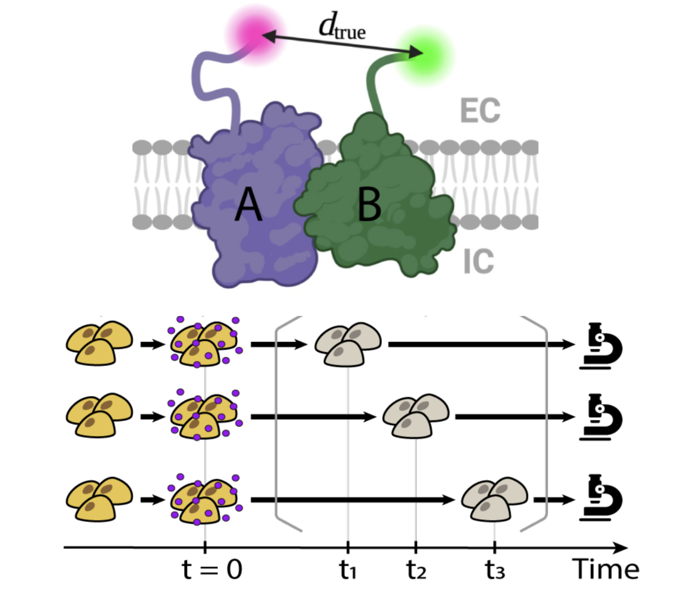
Identifying Intermolecular Interactions in Single-Molecule Localization Microscopy
Xingchi Yan, Polly Y. Yu, Arvind Srinivasan, Sohaib Abdul Rehman, Surabhi Kottigegollahalli Sreenivasa, Jeremy B. Conwaya, Maxim B. Prigozhin†
Abstract
Intermolecular interactions underlie all cellular functions, yet visualizing these interactions at the single-molecule level remains challenging. Single-molecule localization microscopy (SMLM) offers a potential solution. Given a nanoscale map of two putative interaction partners, it should be possible to assign molecules either to the class of coupled pairs or to the class of noncoupled bystanders. Here, we developed a probabilistic algorithm that allows accurate determination of both the absolute number and the proportion of molecules that form coupled pairs. The algorithm calculates interaction probabilities for all possible pairs of localized molecules, selects the most likely interaction set, and corrects for any spurious colocalizations. Benchmarking this approach across a set of simulated molecular localization maps with varying densities (up to ∼55 molecules μm−2) and localization precisions (1 to 50 nm) showed typical errors in the identification of correct pairs of only a few percent. At molecular densities of ∼5 to 10 molecules μm−2 and localization precisions of 20 to 30 nm, which are typical parameters for SMLM imaging, the recall was ∼90%. The algorithm was effective at differentiating between noninteracting and coupled molecules both in simulations and experiments. Finally, it correctly inferred the number of coupled pairs over time in a simulated reaction–diffusion system, enabling determination of the underlying rate constants. The proposed approach promises to enable direct visualization and quantification of intermolecular interactions using SMLM.
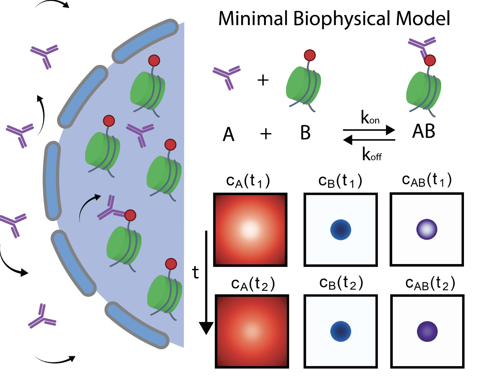
Antibody-trapping presents a widespread pitfall for microscopy and genomics in the nucleus
Konrad Chudzik*, Yuko Sato*, Xingchi Yan*, Simon Ullrich, Watanya Trakarnphornsombat, Lothar Schermelleh, Geoffrey Fudenberg†, Hiroshi Kimura†, Michael I. Robson†, Irina Solovei†
Abstract
Chromatin has a complex 3D structure and diverse binding proteins that coordinate the genome’s most essential functions. Many microscopy and genomics technologies that map chromatin proteins and modifications rely on the diffusion of antibodies (Abs) to target epitopes within whole nuclei. Here, we reveal a critical flaw in such methods that arises when Abs become trapped at the edge of nuclear structures and fail to reach internally positioned epitopes. This “Ab-trapping” results in artifactual peripheral signal that fundamentally distorts the apparent positions of chromatin features across the genome and nucleus. Using computational modeling and experimental validation, we demonstrate that Ab-trapping is caused by a combination of three compounding factors—high epitope abundance, high Ab affinity, and low Ab diffusion rates. Ab-trapping can thus systematically misrepresent the localization of many prevalent chromatin features like histone modifications, transcription factors, nucleolar proteins, and protein tags. We also show that this artifact manifests in multiple technologies, including immunofluorescence microscopy, more recent CUT&Tag-seq, and likely any method relying on Ab diffusion. Finally, we outline readily implementable strategies to identify and mitigate Ab-trapping. Combined, our work presents a previously unrecognized yet prevalent artifact in Ab-based chromatin mapping methods and the means to resolve it.
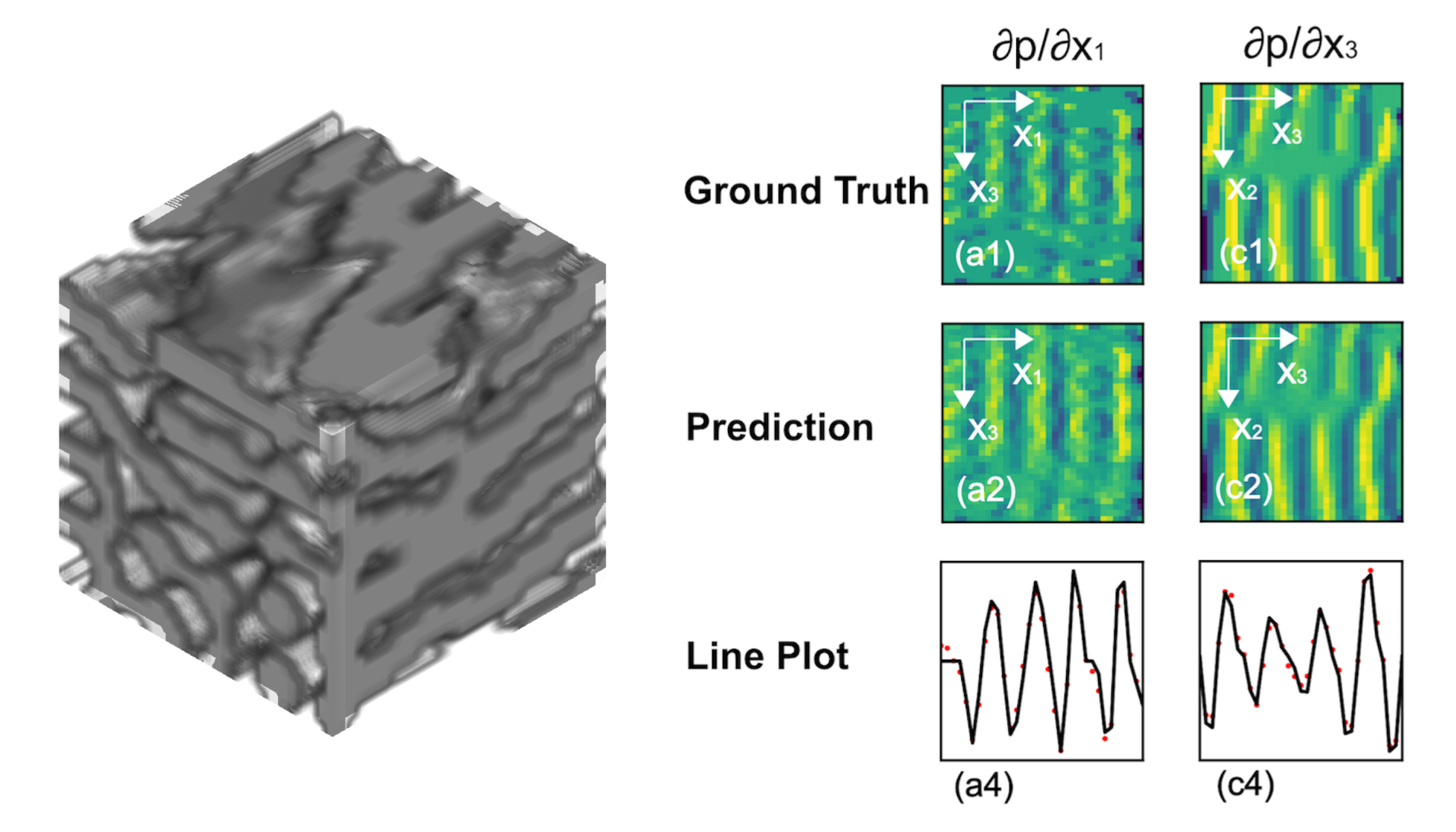
All-optical photoacoustic tomography via beam deflection
Xingchi Yan*†, Siyuan Song*, Hanxun Jin
Abstract
Photoacoustic imaging (PAI) uniquely combines the advantages of optical contrast with deep tissue penetration capability of acoustic waves, enabling imaging at depths of several centimeters. Conventional PAI methods have relied on pulsed lasers to induce the photoacoustic effect, coupled with arrays of pressure transducers to detect the resulting ultrasound signals. In this work, we propose an alternative all-optical approach that leverages optical deflection to record photoacoustic waves by an array of detection beams. The measured signal is shown to be the Radon transform of the pressure gradients. An optimization-based inversion procedure is used to reconstruct the initial time pressure gradient field. Subsequently, a Galerkin method is used to reconstruct the pressure field from the pressure gradient field. The new modality offers the potential for enhanced sensitivity and reduced signal distortion, advancing the capabilities of PAI beyond traditional transducer-based systems.
Determination of Fresnel integrals for X-ray phase contrast imaging with the fast Fourier transform
Xingchi Yan, Gerald J. Diebold†
Photoacoustic effect from an oscillating source in a one-dimensional resonator
Xingchi Yan†, Siyuan Song, Gerald J. Diebold
Abstract
Although the photoacoustic effect is most commonly generated by pulsed or amplitude modulated continuous optical sources, it is possible to generate acoustic waves by moving a constant amplitude, continuous light beam. If the light beam moves at the speed of sound, an amplification effect takes place which can be used in trace gas detection. Here, the properties of the photoacoustic effect are investigated for a continuous optical beam moving in a one-dimensional resonator. The solution shows the additive effects of sweeping the optical beam the length of the cell and back.
Ponderomotive force on an optically levitated sphere in an amplitude-modulated laser beam
Wenyu Bai, Xingchi Yan, Gerald J. Diebold†
Abstract
The motion of an optically levitated, transparent sphere in an amplitude-modulated, Gaussian laser beam is shown to be described by Mathieu functions. Solutions to the equation of motion for the particle are found with and without the effects of viscosity for sinusoidal optical amplitude modulation. Solutions for step function changes in the amplitude of continuous laser irradiation are found as well. By examination of the Hamiltonian field for the particle, it is shown that the particle motion is not harmonic under sinusoidal modulation of the optical beam intensity. With sinusoidal optical modulation, the particle motion shows a steady ponderomotive force to act on the particle proportional to the second space derivative of the laser intensity.
Generation of high amplitude compressions and rarefactions in a photoacoustically excited droplet
Xingchi Yan, Gerald J. Diebold†
Abstract
Photoacoustic excitation of a fluid sphere generates an outgoing ultrasonic wave whose time profile permits determination of the density, sound speed, and diameter of the sphere. Experiments with pulsed laser beams have confirmed the major predictions of existing theory. With regard to acoustic waves generated within spheres, although mathematical expressions for their properties are known, virtually no exploration of the waveforms in theory or experiment has taken place. Here, two cases for photoacoustic excitation of a droplet are discussed: first, absorption of radiation in a region of fluid external to the droplet, and, second, absorption of radiation by the droplet itself. Large amplitude transients, compressions in the former and rarefactions in the latter, are generated as the waves approach the center of the sphere. The high amplitudes of the waves suggest shock wave formation.
Abel inversion of optical beam deflection signals from photoacoustic waves with symmetry in one, two, and three dimensions
Xingchi Yan†, Gerald J. Diebold
Abstract
Photoacoustic waves can be detected by recording optical beam deflection as a result of density gradients inherent in the waves. Beam deflection can be recorded from a single laser beam placed at a distance from the source where data are generated as a function of time, or by recording beam deflections at a single time as the position of the probe beam is varied. For either case, the deflection signal is shown to be described by an Abel transform of the pressure gradient. Here, the inversion of beam deflection data from photoacoustic sources with symmetry in one, two, and three dimensions is investigated. An iterative method based on total variation regularization and a regression procedure based on Lasso regularization are shown to result in accurate reproductions of the theoretical expressions that describe photoacoustic waves from simple sources.
Teaching